
BIO340 Laboratory Guide #8
CONTROL OF HEARTBEAT IN
THE FROG
The beating of a vertebrate heart consists of rhythmic, coordinated contraction (systole) and relaxation (diastole) of the heart's chambers, the atria and ventricles. Each contraction is triggered by an action potential that is initiated in a pacemaker region and spreads to all of the cells of the heart via gap junctions. Because the cells in the pacemaker are modified muscle cells, the vertebrate heartbeat is said to be controlled by a myogenic pacemaker. This characteristic enables the heart to beat independently in the absence of outside influences.
The pacemaker is located in the wall of the right atrium, in a structure called the sinus node, sinoatrial (SA) node (in birds and mammals), or sinus venosus (in fish, amphibians, and reptiles). The pacemaker consists of modified cardiac muscle cells, which have a continuous, moderate conductance to Na+, as well as a class of transient Ca++ channel which opens under hyperpolarizing potentials. This causes a steady depolarization referred to as the pacemaker potential. As a consequence, these cells have no true resting potential; after each action potential, sodium and calcium influxes ramp the membrane potential up and trigger another action potential. This results in rhythmic firing of the pacemaker cells. Excitation initiated in the pacemaker spreads rapidly to the left atrium, resulting in nearly synchronous contraction of both atria (in vertebrates that have two atria), and to the atrioventricular (AV) node, which lies near the junction between the atria and the ventricle(s). From the AV node, excitation spreads through a specialized bundle of conductive fibers, (the bundle of His and radiating Purkinje fibers in mammals) to the apex of the ventricles, and from there throughout the muscle of the ventricles. This causes the ventricles to contract with an efficient wringing action.
Nodal cardiac cells are capable of displaying their own endogenous pacemaker activity, even when isolated from the rest of the heart. However, it is the cells in the sinus node which have the highest intrinsic rate of firing. Because all cardiac cells are coupled by gap junctions (electrical synapses), depolarization spreading from the sinus node causes cells in other parts of the heart to contract sooner than they would in response to their own internal rhythms. Consequently all cardiac cells contract at the same rate as the pacemaker in the sinus node, but with differing phase relationships.
The intrinsic, myogenic rhythmicity of the heart can be modulated by both neuronal and hormonal input. Parasympathetic preganglionic axons in the bilaterally paired vagus (tenth cranial) nerves synapse in a ganglion situated within the wall of the heart, close to the atria. Postganglionic parasympathetic neurons slow the heartbeat by releasing acetylcholine (ACh) onto the pacemaker cells. In contrast, the heart rate can be accelerated by norepinephrine released from postganglionic sympathetic neurons, or by epinephrine released by the adrenal medulla.
This laboratory exercise will give you the opportunity to examine some of the factors which influence the heartbeat of the turtle. While the ventricle in a mammalian heart is completely divided into separate right and left chambers, the turtle heart has a ventricle that is only partially divided. This anatomical feature results in some exchange of oxygenated and deoxygenated blood between the pulmonary and systemic circulations. The relative flow of blood to the lungs and to the rest of the body is determined by the resistance along each pathway. When the turtle breathes, blood flow to the lungs is high, but when the turtle halts breathing during a dive, blood is shunted from the lungs to the rest of the body. Except for this difference, the turtle heart is quite similar to mammalian hearts.
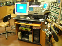
I. EQUIPMENT SETUP
As in previous experiments, you should have all of your equipment set up and tested before you begin the dissection. The muscular contractions of the heart will be monitored with a different kind of displacement transducer than that used to measure contractions of the frog gastrocnemius. In this experiment, the recordings will be made with the heart left in the animal (in situ). Contractions of the ventricle and one of the atria will be monitored by using both force and diplacement transducers. Two stimulators will be used: the SD9 to stimulate one of the vagus nerves, and the built-in PowerLab stimulator to directly excite the ventricle.
The electronic equipment needed for this experiment includes an electronic stimulator, two stimulating clips, a stimulating probe, a force transducer, two displacement transducers, heart hooks and clips on threads, an ETH-400 transducer bridge amplifier, and a PowerLab/PC station.
Equipment Setup Procedure:
1) Connect the lead of one of the two horizontally-mounted displacement transducers to the Channel 1 input of the ETH-400 bridge. Connect the lead of the vertically-mounted force transducer to the Channel 2 input of the ETH-400. For now, leave the lead of the second displacement transducer unattached. Connect the corresponding ETH-400 outputs to the input jacks of CH1 and CH2 on the PowerLab box.
2) Turn on the PowerLab box, boot the PC, and launch Chart. Make sure that the first four channels are turned on. For now, expand the display areas so that Channels 5-8 are completely compressed and Channels 1 and 2 occupy about three times as much space as Channels 3 and 4.
3) Set the sampling rate (upper right) at 1K/sec and the horizontal plotting compression ratio (lower right) at 20:1.
4) Set the Range: for channels 1 and 2 at 1 V and the Range: for channels 3 and 4 at 5 V. Set the Low Pass filter to 50 Hz in the Input Amplifier... window for Channels 1 and 2. Set all four channels for monopolar (positive input only) recording. Use the Display Offset... window and the ETH-400 offset knobs to adjust channels 1 and 2 to read approximately 0 Volts.
5) Use Channel Titles under the Display menu to label the four recording channels as "Atrium", "Ventricle", "Vagus Stim", and "Vent Stim", respectively.
6) Check out your setup with the instructor before proceeding. Make sure all of your equipment is set up properly and that you know the locations and functions of all of the controls. Refer to the tutorial for Lab 1 or consult the instructor if you have any questions.
II. HEART AND VAGUS NERVE EXPOSURE AND
PREPARATION FOR RECORDING
Work rapidly, but carefully during dissections, and always read through the entire procedure before you begin. Wear gloves. Be especially careful when exposing the vagus nerves and when applying clips to the heart.
Your laboratory instructor will provide your group with a bullfrog that has been cold anesthetized and has had its brain and spinal cord pithed.
Dissection Procedure and Initial Transducer Attachment:
1) Center the frog on its back in a rubber-lined dissecting pan. Firmly pin it in place with crossed large T-pins through each of the forearms and each of the thighs, stretching the body between them. It is essential for the first part of this lab that the trunk of the frog not be free to move relative to the pan. Secure the pan in place to the table with four small lumps of clay.
2) Make a midline ventral incision along the entire length of the abdomen, thorax, and neck, and retract the skin on both sides.
3) Use scissors to carefully make a shallow midline incision through the ribcage, up to the base of the neck. Spread the incision open and locate the heart, as well as the two vagus nerves leading from the heart into the neck. Identify the three chambers of the heart.
4) Make lateral cuts through the rib cage just wide enough to allow free access to all three chambers of the heart. Completely remove the cut sections of the rib cage.
5) Gently free each vagus nerve from the neighboring blood vessels and connective tissue, using the glass rods to manipulate the nerves. Pass a thread under both nerves, so that they can be lifted onto the stimulating electrodes at a later time. Do not cut the nerves and avoid pulling on them any harder than absolutely necessary. Place a KimWipe saturated with Ringer's solution over the area to keep it from drying out.
6) Carefully lift and slit the pericardium to expose the heart.
7) Attach the small heart hook connected to a thread to the tip of the ventricle. Be sure to pass the hook through only the muscular tip of the ventricle and not through the ventricular chamber. Connect the thread to the vertical Channel 2 force transducer lever. Angle the transducer, and position its stand so that the ventricle is held clear of the underlying tissue with no slack in the thread and minimal tension on the heart between beats. This transducer will be used to monitor contraction strength of the ventricle.
8) Use extreme caution for this next step. Connect the right atrium (frog's right, NOT your right) to a small heart clip attached to another thread. Try to grab just the outer layer of tissue of the atrium. If the jaws of the clip penetrate the thin walls of the atrium, the pericardial cavity will quickly fill with blood and the experiment may have to be terminated. Do not attach the other end of the thread at this time.
9) Carefully remove any clotted blood from the pericardial cavity and flush it with Ringer's.
III. RECORDING CARDIAC MUSCLE TENSION
As you work through the following procedures, be sure to save important Chart records to disk. Keep track of your chart records by making Comments. Create a folder for your group on the hard disk, and save your file periodically. Rename your file periodically using the Save As... option, so that older versions of your file are maintained (so if you mess up you don't lose all of your data). If you are instructed to print out a Chart record, do that after the experiment, while you are preparing your data sheet. Use your time during the experiment to collect the data, making sure you have recorded, saved, and annotated the records you will need for the print-outs and measurements you will make later.
1) Confirm that the force transducer and its stand are positioned so that the ventricle is held clear of the underlying body wall, that there is no slack in the thread, and that there is minimum tension on the thread between beats.
2) Start the Chart recording. Only Channel 2 should show an active trace. Adjust the ETH-400 bridge to approximately zero the trace between beats. Adjust the channel zero and range so that the full excursion of the ventricular contraction takes up approximately 1/2 of the available channel display range. If necessary, reverse the channel polarity so that contractions of the heart chambers produce upward deflections of the recording traces. You may see a bump riding the leading edge of each ventricular contraction. This is an artifact of the movement of the atria, just preceding each ventricular contraction.
3) Record at least 20 seconds of activity, then hit the tiny graph next to the START/STOP button in order to ceasing saving the recorded trace. Carefully slide the transducer stand ~2 mm further from the frog, to stretch the heart muscle. Re-zero the trace using the ETH-400 zeroing knob. Hit the tiny graph button again to begin saving another 20 seconds of activity.
4) Repeat step 4 at least 5 more times, or until it does not seem possible to stretch the heart any further without damaging it.
5) Slide the transducer stand back towards the frog to relieve the tension on the heart.
6) Label each of your recorded Chart segments with the length of stretching applied to the ventricle.
Q1: What is the expected relationship between length and tension for cardiac muscle? Is it exactly the same as for skeletal muscle? Why or why not? How does this length/tension curve relate to "Starling's Law of the Heart"?
IV. RECORDING AND ALTERING THE CARDIAC CYCLE
A. Restting Transducers
1) Detach the ventricular hook thread from the force transducer and slide the transducer stand out of the way.
2) Unplug the force transducer lead from Channel 2 of the ETH-400 and replace it with the lead of the remaining displacement transducer
3) Position the two displacement transducer stands so that the end of each transducer lever is directly over the heart.
4) Carefully attach the atrial clip thread to the lever of the Channel 1 and the ventricular hook thread to the lever of the Channel 2 displacement transducer. Carefully adjust the position of each transducer so that it moves cleanly in response to its respective heart chamber.
B. Normal Cardiac Activity
1) On Chart, adjust the ranges for Channels 1 and 2, shift/stretch the vertical axes, and change the horizontal compression ratio, if necessary, so that the contractions of the ventricle and atrium can be seen clearly. You may also see the contraction of the sinus venosus as a small bump or inflection in the leading edge of each atrial beat. Similarly, you may see an artifact of the atrial contraction on the ventricular trace.
2) If necessary, reverse the channel polarity so that contractions of the heart chambers produce upward deflections of the recording traces.
3) Record at least 60 seconds of stable baseline activity.
Q2: Are the atrium, ventricle, and sinus venosus beating at the same rate? If so, what is the approximate phase relationship between the contractions of these three structures (e.g. 0� is synchrony, 90� is a quarter cycle lag, 180� is exactly out of phase)?
B. Effect of Vagal Stimulation on Heartbeat
1) Detach the force transducer from the third ring stand and mount a stimulating probe with two silver hooks.
2) Uncover the vagus nerves and gently lift both onto the stimulating electrodes. Moisten the nerves with Ringer's solution, then cover them with a layer of Vaseline to keep them from drying out. Be careful not to bump the probe that holds these electrodes, since you might stretch and damage the nerves.
3) Using appropriate cables connect the SD9 stimulator output jacks to the stimulus probe and to the + input jack of the PowerLab CH 3. This later set of connections will allow you to record the pulses that you use to stimulate the vagus nerve.
4) Set up channel 3 on Chart for monopolar recording with no filters, and set the Range: to 1 volt.
5) MAKE SURE THAT THE STIMULATOR MODE IS SET TO OFF. Turn on the stimulator and set it up for 0.1 V x 6 msec pulses at a frequency of 1 Hz.
6) While recording the ongoing heartbeat, stimulate the nerve with 5 second trains of pulses. Start with an amplitude of .1V and, if there is no effect on the heart rate, first increase the frequency to 5Hz, then gradually increase the stimulus voltage until you see an effect. DO NOT EXCEED 10 VOLTS ON THE STIMULATOR. You may have to adjust the range on Channel 3 to keep the recorded pulse from going off scale. If you fail to see an effect, consult with the instructor.
7) Use a series of 5 second trains of increasingly intense stimuli to determine the threshold for partial inhibition of the heartbeat (where the heart rate slows) and for complete inhibition (where the heart stops beating altogether). Stop stimulating as soon as the heart stops beating. Allow the heart to recover to its normal baseline rate between tests.
8) Allow Chart to run while you stimulate and look for partial and complete inhibition. Document on a separate sheet of paper all stimulator settings and the corresponding elapsed times from the Chart record. Make sure that you STOP stimulating when you are finished.
9) When you are finished stimulating, transfer your recorded stimulation parameters to the Chart record at the appropriate locations.
Print out a well-labeled record showing partial inhibition and complete inhibition, and indicate the stimulus settings that were used to produce each level of inhibition. On this record, measure the following parameters for each level of inhibition, all of which (except the recovery period) should be clearly visible on your print-out:
a. the stimulus intensity settings for partial and complete vagal inhibitionb. the atrio-ventriculr (A-V) interval during each
c. the amplitude of contractions, if these differ from baseline (OK to express as mV)
d. the recovery periods(time taken for recovery of the original rate and amplitudeof contractions, from the end of the stimulation train) from partial and complete
inhibition
Q3: Explain any differences between the normal heartbeat and the heartbeat during partial inhibition.
Q4: During complete inhibition, is the heart arrested in systole or diastole? When the heart recovers from complete inhibition, which chamber(s) contract first?
Q5: Which chambers of the heart contract during escape from vagal inhibition? Explain.
C. Ventricular Refractory Period
1) Connect the positive stimulus output jack on the PowerLab stimulator to both the channel 4 + input jack and to the exposed chest muscle, using a stimulating clip.
2 Carefully, attach a second stimulating clip near the tip of the ventricle and connect its lead to the negative stimulus output jack of the PowerLab. This clamp is sharp, so be very careful not to puncture the ventricle.
3) Set Channel 4 to unfiltered monopolar recording with a range of 5 volts. This setup will allow you to record the pulses that you use to stimulate the ventricle.
4) Set up the PowerLab stimulator for continuous pulses at 0 V x 6 msec x 0.2 Hz and set the stimulator amplitude range to 2 V. Activate the Stimulator Panel, so you can adjust the stimulus parameters while recording. Set the Increment to .1 Hz. To do this, hold down the command key on the keyboard and simultaneously press the duration button in the stimulus panel. A dialog box should open, which will allow you to reset the duration increment down to 1 msec.
5) Start the Chart recording. Use the vagal nerve stimulation to produce complete inhibition, and then stimulate the ventricle. Deliver pulses to the ventricle at 0 V x 6 msec x 0.2 Hz. Increase the stimulation voltage until each pulse causes a ventricular contraction.
6) When you reach the threshold for ventricular contraction, slowly increase the stimulation frequency. Continue to increase the stimulation frequency until you reach a point where ventricular contractions no longer follow the stimuli 1-for-1. The reciprocal of the stimulation frequency at this point is the refractory period of the ventricle.
7) Turn off both the vagal and ventricular stimulation and allow the heart to recover its normal cycle.
8) Repeat steps 5-7 at least twice, saving and labeling your Chart records as you go. Turn off both vagal and wentricular stimulation when you are finished and allow the heart to recover its normal cycle.
Alternatively - You can quantify the ventricular refractory period through a careful study of the period following each ventricular contraction during which it is impossible to produce an EVS (see below). Print out well labeled records quantifying the refractory period by this method.
Q6: Explain the rationale for using this method to determine the ventricular refractory period.
Q7: Of what value is the refractory period to the normal functioning of the heart? In your explanation, be sure that you clearly distinguish between cardiac excitation and cardiac contraction.
D. Ventricular Extrasystoles
A ventricular extrasystole is a premature ventricular excitation and contraction that originates in the ventricular conduction fibers and is not preceded by atrial excitation and contraction. Spontaneous ventricular extrasystoles can seriously disrupt the normal rhythmic cardiac cycle.
1) Start by adjusting the electronic stimulator settings to match the PowerLab settings from the previous study.
2) Uncouple the PowerLab box from the electronic stimulator synch input. Unplug the vagus nerve stimulating leads from the electronic stimulator. Unplug the ventricular stimulating leads from the PowerLab box and plug them into the electronic stimulator outputs. Make sure that the lead running to the ventricle is the negative one.
3) Deliver single pulses from the electronic stimulator to the ventricle. Time your pulses so that they arrive at different times in the ongoing ventricular beat cycle. If your stimulus parameters are set correctly and you stimulate at an appropriate time, you should be able to generate "extra" beats in the ventricle.
4) Save your labeled Chart record when you are finished.
Q8: Is there any phase of the cycle when extrasystoles cannot be produced? If so, can you relate this time to the ventricular refractory period which you measured above?
Q9: Is there any effect of ventricular extrasystoles on the atrial rhythm? When an extrasystole is produced, how is the timing of subsequent ventricular contractions affected (advanced/delayed/eliminated)?
E. Mini- Independent Study on the Pharmacology of Autonomic Control
Using the supplied acetylcholine chloride (parasympathetic neuromodulator), carbamyl choline chloride (ACh agonist), atropine (ACh antagonist), and/or norepinephrine (sympathetic neuromodulator) design, conduct, and record an experiment on pharmacological control of the heartbeat.
OR
Carefully string a loop of tread around the heart between the atria and ventricle. Slowly tighten the thread and see if you can denmonstrate either partial (1st or 2nd degree) or complete (3rd degree) heart block - a dissociation of the beats of the atria and ventricle.
F. Shutting Down
1) Make sure that you have saved all of your data to the hard drive, then quit Scope. Turn off both the PowerLab box and the stimulator. Disconnect the cables from the stimulator, the PowerLab, and the turtle.
2) Arrange with the instructor to properly dispose of the remains of the frog. Clean and dry all of your surgical instruments. Rinse out the dissecting pan.
3) Return all chemical solutions to the refrigerator.
IV. PREPARATION OF THE LAB DATA SHEET
Your data sheet should include at least FOUR of the FIVE items described in the boxes above. Make sure that the axes of all of the graphs and print-outs are labeled and calibrated. You should certainly discuss your results and the answers to the questions with your partners and others in the lab. However, please work independently when you prepare your data sheet.