
BIO325 Laboratory Guide #9 (2024)
ACTION POTENTIALS I:
ELECTRONIC SIMULATIONS OF ACTION POTENTIALS
falls under category
In this lab you will investigate the "active" membrane properties involved in producing neuronal action potentials. The gated equivalent circuit model (GECM) has been built according to the standard, Hodgkin-Huxley "equivalent circuit" model of the neuronal membrane. With this model you control the timing of state-changes (openings and closings) of gates which control ion flow through the ion-specific channels of the membrane. The model is capable of producing a reasonable facsimile of an action potential, slowed by a factor of 1000. The point of this simulation is to give you a real "hands-on" feel for the sequence of gating and conductance changes underlying the action potential.
To get the most from this simulation, don't read too far ahead. Conduct the first part of the simulation (Section I) as an exploratory exercise - using the simple process of trial and error learn the mechanics of how to produce an action potential. Then use the second part (Section II) to refine your understanding of the equivalent circuit concept, how this concept is realized in the model, and what aspects of the neuronal membrane are omitted or oversimplified in this model.
Photo of Model
I. GATED EQUIVALENT CIRCUIT MODEL - EXPERIMENT
A. Initial Setup
1) Start up the PC (if necessary), turn on the PowerLab box, and open the AP Template file. This will launch the Scope application, make appropriate PowerLab settings, and load a set of sample traces.
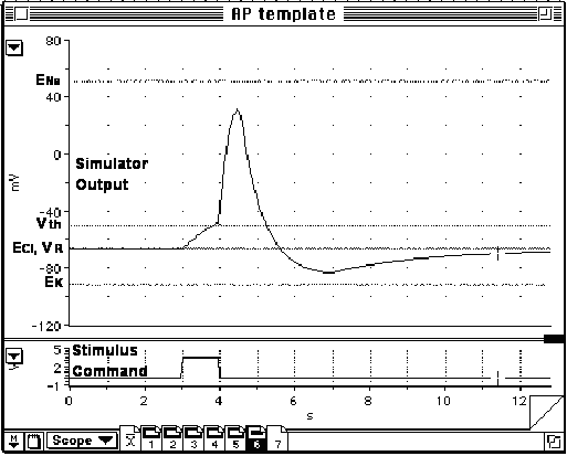
2) Examine the PowerLab display, which should appear similar to the sample in the link to the right. The upper trace displays Input A. A "template" action potential which you are to attempt to match appears in red. Additional lines correspond to the equilibrium potentials for sodium (ENa), potassium (EK), and chloride (ECl), as well as the resting potential (VR) and the threshold potential (Vth). The lower trace displays Input B. The output pulse of the PowerLab stimulator appears in blue.
3) Under the Setup / Sampling menu verify that Mode is set to Repetitve.
4) Connect two screws on the gated equivalent circuit model (GECM) board to the CH1 input of the PowerLab using a BNC-to-double banana plug cable and two alligator clip adapters. Make sure that the "IN(SIDE)" screw on the GECM is connected to the active (nontab) banana plug and the "OUT(SIDE)" screw on the GECM is connected to the common or ground (tab) banana plug.
5) Plug all three power supplies into the GECM, matching them by color (green, red, and yellow).
6) Turn on the power strip(s) for the power supplies.
7) Connect the PowerLab Output+ to the CH2 input via a BNC cable and T-connector. Connect the free end of the T-connector to the white stimulus input on the GECM via a BNC cable, BNC-phono plug adapter, and phono plug to 1/8" phone plug adapter.
8) Set all three switches on the GECM to the off (down) position.
9) Start a continuous series of PowerLab sweeps via the Start button on the Scope display. The new red membrane potential trace should show a constant value corresponding to the resting potential VR of about -70 mV, with a rounded upwards deflection at the time of the stimulus pulse. The new blue stimulus trace should match the template stimulus trace with a square upwards deflection. You should also hear a distinct "click-click" produced by a magnetic relay on the GECM board. If these initial conditions are not met, consult with the instructor.
10) Disconnect the stimulus cable from the GECM.
B. A "Blind" Experiment
You have three toggle switches and one push-button switch which you can manipulate on the GECM. These are labeled A, B, C, and D. Your task for this part of the laboratory exercise is fairly simple:
1) Experiment with throwing switches A, B, and C and pushing button D as Scope repetitively sweeps out new traces on the computer screen.
2) Continue until you have determined a set of starting switch positions, a sequence of switch changes, and a timing of switch changes which results in a trace closely matching the template action potential. All switches should be returned to their initial positions by the end of the trace.
3) Once you think that you have the correct timing, reconnect the stimulus cable to the GECM via the 1/8 inch phone plug adapter. Now the Scope software will produce the stimulus pulse and you just have the three toggle switches to worry about.
Q1: When you think you have a solution which closely matches the template, call the
instructor over to demonstrate your solution.
If you are having trouble figuring out how accomplish this, here is a systematic way to proceed:
Step 1: Determine the effect of each switch on the membrane potential and the time-course of each such effect. For each toggle switch up (towards the circuit board) is on and down (away from the circuit board) is off. For the push-button switch the depressed position is on and the released position is off. Note: Not all of the switches have independent effects - the effect of turning on one switch may depend on whether another switch is on or off.
Step 2: Break the action potential template into components. Determine what switch and/or button manipulations are required to produce each component. For example:
What switch setting(s) produce(s) the resting potential VR of about -70mV?
What produces the initial stimulus hump (at 5.0-5.7 seconds)?
What produces the rising, depolarizing phase (at 5.7-6.0 seconds)?
What produces the falling, repolarizing phase(at 6-7 seconds) and
undershoot, hyperpolarization phase (at 7-8 seconds)?
What produces the recovery from undershoot (at 8-11 seconds)?
How and when during the course of the action potential can each switch be
restored to its starting position?
Step 3: When you have duplicated the rough shape of the action potential template, work on refining your timing to provide a closer match. Refine your technique to get the simplest series of switch manipulations which reliably produces an action potential template match.
Stop reading here until you have successfully completed sections A and B.
II. GATED EQUIVALENT CIRCUIT MODEL - COMPONENTS
A. The Membrane Equivalent Circuit
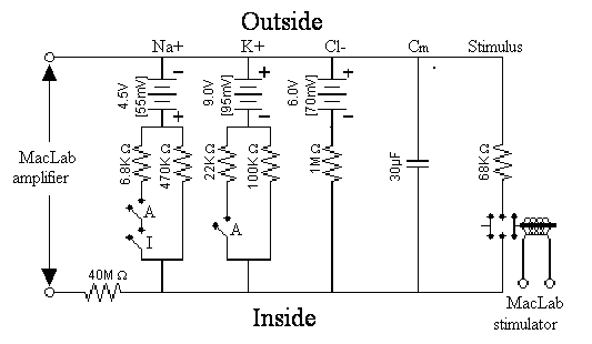
Examine the circuit diagram for the GECM with which you have been experimenting, by clicking on this link. Work with this diagram until you thoroughly understand what each component represents in an actual neuron membrane. You should be able to identify each of the electronic components on the circuit board. We will be exploring the equivalent circuit model at length in class, so bring your questions with you.
The GECM model has six small circuits, arranged in parallel, which model specific components of the neuronal membrane - specifically a space-clamped axon. Working from left to right in this diagram and on the GECM circuit board:
1) Recording - This consists of the PowerLab amplifier, connected across the modeled neuronal membrane (the two screws labeled �OUT� and �IN� at the extreme right end of the GECM board). By convention, transmembrane potentials are recorded inside relative to outside, hence the active (positive) input of the PowerLab is connected to the inside of the simulated neuron and the common (negative, ground) input to the outside. A 40 MW series input impedance, produced by four 10 MW resistors (brown-black-blue bands; diagonally mounted at the lower left end of the circuit board in the active recording lead), functions as a voltage divider. This insures that the PowerLab drains negligible current off the rest of the circuit and does not itself affect the measured membrane potential. It also scales down the measured voltage into the appropriate millivolt range. An additional recalibration of the recorded potential is performed by the PowerLab Scope software.
2) Sodium - According to Ohm's Law for ionic currents, sodium is pushed across the membrane by a driving force equal to the membrane potential minus the sodium equilibrium potential (Vm - ENa). In this model, ENa is set to approximately +55 mV by a +4.5 V voltage supply (green phone jack). Sodium current flows through selective membrane channels, represented in this model by two routes:
a) The voltage-gated, active sodium conductance is represented by a 6.8 KW resistor (blue-gray-red bands) wired in series with the first two switches, labeled "A" and "B", on the board. These two switches, marked "A" and "I" in the diagram above, represent the sodium "activation" and "inactivation" gates of the membrane sodium channels. Both switches must simultaneously be in the "on" position in order for current to flow through this low conductance route.
b) The resting sodium conductance through these same channels is represented by a separate route through a 470 KW resistor (yellow-purple-yellow bands).
Current flowing through the combined sodium conductance pulls the measured membrane potential towards the sodium equilibrium potential ENa (approx. + 55 mV).
3) Potassium - Potassium is pushed across the membrane by a driving force equal to the membrane potential minus the potassium equilibrium potential (Vm - EK). In this model, EK is set to approximately -90 mV by -9.0 V voltage supply (red phone jack). Potassium current also flows through selective membrane channels, represented in this model by two routes:
a) The voltage-gated, active potassium conductance is represented by a 22 KW resistor (red-red-orange bands) wired in series with the third switch, labeled "C" on the board. This switch, marked "A" in the diagram above, represents the potassium "activation" gates of the active membrane potassium channels. This switch must be in the "on" position in order for current to flow through this low conductance route.
b) The resting potassium conductance through these same channels is represented by a separate route through a 100 KW resistor (brown-black-yellow bands).
Current flowing through the combined potassium conductance routes pulls the measured membrane potential towards the potassium equilibrium potential EK (approx. -90 mV).
4) Chloride - Chloride is pushed across the membrane by a driving force equal to the membrane potential minus the chloride equilibrium potential (Vm - ECl). In this model, ECl is set to approximately -70 mV by a -6.0 V voltage supply (yellow phone jack). Chloride current can flow through the membrane only through a small constant chloride conductance, represented by a 1 MW resistor (brown-black-green bands). This voltage-gated conductance pulls the measured membrane potential towards the chloride equilibrium potential ECl (approx. -70 mV) which is approximately equal to the resting potential VR. In your particular model, ECl may be anywhere in the Vrest + or - 5mV range
5) Membrane Capacitance - As discussed in earlier labs, the lipid bilayer is a nonconductor situated between two conductive mediums, the intracellular and extracellular spaces. Because of this, dissimilar charges can accumulate on the two sides of the membrane, held loosely in place by their electrostatic attraction across the membrane. This property of separating charge is called capacitance. The membrane capacitance for this model is set to 30 mF and is produced by three 10 mF capacitors (blue cylinders) wired in parallel. This capacitance value was chosen to produce model kinetics (rates of potential change) which are about 1000 times slower than those of an actual neuronal membrane. These capacitors "round out" the shape of the action potential. If these capacitors are removed from the circuit any change in membrane conductance produced by throwing a switch results in an instantaneous membrane voltage change.
6) Stimulation - Action potentials are normally triggered by transient depolarizations of the neuronal membrane. In this model stimulus depolarizations are produced by closing either one of two switches which temporarily short the inside and outside of the membrane together through a 68 KW resistor (blue-gray-orange bands) located at the upper right of the circuit board. This actually corresponds well to an excitatory postsynaptic potential produced by opening up a non-selective monovalent cation channel, such as that associated with the nicotinic acetylcholine receptor or with the fast glutamate receptor. In the model this stimulating circuit may be manually activated via the red push-button switch labeled "D" or automatically via the magnetic relay which is controlled by the PowerLab stimulator output (white phone jack).
B. Voltage-Gated Conductances
In this simulation you control the switch "gates", which in turn control the relative conductances to sodium and potassium and produces the current fluxes and membrane potential changes associated with the action potential. However, in an actual neuron, these gates are controlled by the membrane potential itself, in specific time-dependent manners. Before you leave this simulation it is important that you reconcile your AP solution with the following "rules" for gate (switch) states and kinetics.
Note: The terminology for switches vs. gates is confusing - an ion channel gate conducts when it is open while an electrical switch conducts when it is closed. Thus closing a switch to the "on" position models opening the corresponding gate, while opening a switch to the "off" position models closing the corresponding gate. For the discussion below we will use "on" and "off" to refer to switch states and "open" and "closed" to refer to the corresponding gate states.
1) Sodium Activation - At rest this switch should be off, (deactivated and closed gate) blocking conductance through this route. It turns on (activates and opens) rapidly in response to a suprathreshold depolarization, such as that provided by the stimulus pulse. It turns off (deactivates and recloses) rapidly when the membrane repolarizes towards the resting potential or below.
2) Sodium Inactivation - At rest this switch should be on (deinactivated and open gate). It turns off (inactivates and closes) slowly as the membrane depolarizes. It turns on (deinactivates and reopens) slowly as the membrane repolarizes back toward the resting potential or below. Very Important Note: Although deactivation and inactivation will both close a sodium channel, they are NOT the same thing. Deactivation and inactivation refer to two separate processes governed by two structurally separate gates with very different voltage dependencies and kinetics (speeds of action).
3) Potassium Activation - At rest this switch should be turned off (deactivated and closed gate. It turns on (activates and opens) slowly as the membrane depolarizes. It turns off (deactivates and recloses) slowly as the membrane repolarizes back toward the resting potential or below.
At this time you should rerun and perhaps modify your action potential solution so that it conforms to these rules.
Q2: Demonstrate your new solution to the instructor.
Data Sheet Item #1:
Use the screen capture utility to produce a printout of your simulated action potential superimposed on the template AP. Clearly mark on your trace the starting state of each switch and when and how each switch was manipulated.
C. Equilibrium, Resting, and Threshold Potentials
A good deal of trial-and-error manipulations were performed in originally designing the GECM and determining "appropriate" values for its components. As you have no doubt noticed, the action potential "template" is actually the sixth in a series of six traces. The previous five traces correspond to the sodium, potassium, and chloride equilibrium potentials, the resting potential, and the threshold potential for initiating an action potential.
Q3: How do you suppose each of these traces was produced? Physically manipulate the model by resetting switches, disconnecting power supplies, and/or adjusting power supply voltages as necessary to duplicate each of these traces. Be sure that you restore the GECM board to its original configuration when you are finished.
Ionic equilibrium potentials in the model are determined by the settings on the three DC power supplies.
Q4: What would be the expected effect on the resting potential of changing the equilibrium potential for sodium? Of potassium? Of chloride? Verify your predictions by changing the appropriate power supply setting(s). Return the power supplies to their original settings when you are finished.
The membrane capacitance in the model is supplied by the three 10 mF capacitors.
Q5: What would be the expected effect on the AP waveform of increasing or reducing the membrane capacitance? How might you test this by adding or removing model components?
Minimal and peak voltage-gated channel conductances are determined by the resistors in the model.
Q6: What would be the expected effect on the resting potential and/or the action potential waveform of increasing or decreasing the minimal sodium conductance? Maximal sodium conductance? Minimal potassium conductance? Maximal potassium conductance? Chloride conductance?
Q7: Myelinated axons have virtually no voltage-gated potassium channels at their nodes of Ranvier. What would be the expected result of omitting this conductance on the shape of the action potential? What conductances/currents repolarize the axon under this condition? Would the action potential have a hyperpolarizing "undershoot?
Please return the model to its original configuration when you are finished with this section.
D. Gate Populations and Kinetics - What is Wrong with this Model
Although this model produces a reasonable slow-motion facsimile of an action potential, it incorporates several simplifications and one deliberate misrepresentation. In particular, the simple two-state switches vastly simplify the actual gating situation. Computer simulations of action potentials, such as that comprising the second half of this exercise, can be used to effectively highlight some of these theoretical aspects of active ion channels and simulating experimental determinations of the voltage- and time- dependence of ion channel gates. For now you should recognize the following:
1) Flow of either sodium or potassium through the membrane occurs via a population of independent ion-selective channels. At any point in time these individual macromolecular channels and their constituent gates are not all in the same state. Thus, cation-specific conductances which we model here as single on/off, open/closed pathways are actually graded in a voltage- and time-dependent manner in a real neuron. Furthermore, the resting conductances for sodium and potassium are not quite the minimal values that occur during an action potential. The Neurons in Action exercises in the next lab will introduce you to these dependencies and allow you will isolate and study both of these dependencies in the future voltage-clamp simulation. Replacing the three switches in our model with potentiometers (variable resistor knobs or siders) would provide a more accurate representation, but would make the model prohibitively difficult to operate for anyone with only two hands. If you have time, you can test such a model, which the instructor has built. It is extraordinarily challenging to "drive".
2) To accurately match their squid giant axon data, Hodgkin and Huxley (H-H) came up with a set of equations, including the following equations for calculating instantaneous sodium and potassium conductances (gNa and gK):
gNa = GNam3h gK = GKn4 where Gx = maximal conductance g for ions Na or K
These equations model the sodium activation gate as having three independent components (indicated by the H-H m3 term). All three of these, in addition to the single inactivation component (the H-H h term), must be simultaneously open in order for that channel to pass ions. Similarly each potassium activation gate has four independent components (indicated by the H-H n4) term, all of which must be simultaneously open in order for that channel to pass ions. Each of these terms is actually a continuous value between 0 and 1 which represents the proportion of open gates at any moment in time. Needless to say, replacing our three switches with eight potentiometers would make our model even more difficult to operate.
3) As indicated, the switches by themselves would produce a disturbingly squared-off action potential. The "curviness" of a real action potential is due in large part to the active population kinetics of these populations of channels, and not just the passive membrane capacitance as in our model. Indeed, in order to smooth out the simulated action potentials and slow the kinetics, the transmembrane capacitance of the model is way out of proportion to that in the real neuron. In other words, this model is a good way to learn about relative timing of voltage-gated conductance changes, but a rather misleading way to learn about the factors determining the actual shape of an action potential.
4) For H-H the gates were hypothetical entities and the sodium activation and inactivation gates were modeled as being independent of each other. The Reichert text takes a more modern view in modeling the sodium channel as having three primary conformations - "closed", "open", and "inactivated", rather than entirely independent "activation" and "inactivation gates. This reflects the fact that the activation and inactivation gating mechanisms of a single channel are actually conformationally-interdependent subunits of a single large macromolecular Na+ channel complex.
Q8: How could you redesign the Gated Equivalent Circuit Model board to match the model presented in Reichert?
E. Shutting down the electronic simulation
1) Exit Scope and turn off the PowerLab box.
2) Turn off the powerstrip(s) powering the electronic circuit board and unplug all of the cables.
II. PREPARATION OF THE LAB DATA SHEET
Your data sheet should include BOTH of the items described in the boxes above. Make sure that the axes of all of the graphs and print-outs are labeled and calibrated. You should certainly discuss your results and the answers to the questions with your partners and others in the lab. However, please work independently when you prepare your data sheet.
The writeup
for this lab
falls under category
B