Resistor/Capacitor
Cable Properties Model
Board A Board B Board C
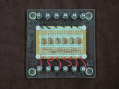
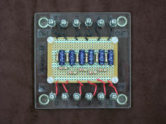
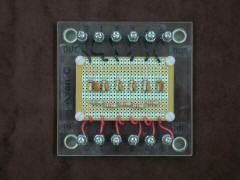
This is a set of three circuit boards, each representing a short length of axon as a set of six identical cylindrical compartments. Each compartment is represented by a high transmembrane resistance Rm and capacitance Cm. Each compartment is separated from its neighbors by lower intracellular logitudinal resistances Ri. The extracellular longitudinal resistances are assumed to be insignificant, hence the extracellular sides of all compartments are simply shorted together to a common extracellular space. Recording access to each compartment is provided by a pair of simple machine screw posts - intracellular (red) and extracellular (black).
As detailed in the diagram below, board A models a small diameter axon. Board B models a large diameter axon, 10x the diameter of A, with a correspondingly increased Cm (10x) and decreased Rm (.1x) and Ri (.01x). Board C models a small diameter axon, but with the middle four compartments myelinated. The outer two compartments and all intracellular resistances Ri match board A, but in the middle four myelinated compartments Cm is decreased (.01x) and Rm is increased (100x).
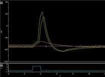
As described in Laboratory #8 of the BIO325 manual, this model is used with a square-wave stimulus pulse (supplied by an electronic stimulator) to study both capacitive rounding and spatial amplitude decay with distance of a transient input signal. This introduces the student to the concepts of the time constant tau for capacitive rounding and the space constant lambda for distance decay. Students also discover that the large diameter axon involves significantly greater current flow (i.e. metabolic cost) than either small axon. Finally, students compare rise time to threshold at a recording site (node 5) distant to the stimulating site (node 0) across the three axon models. This will ultimately be related to increased axon diameter and myelination as alternative ways to speed action potential propagation.
Printouts of the results of using this model as described in laboratory #8 are available online in a poster (PowerPoint format) or upon request from brhoades@wesleyancollege.edu .